
心エコー
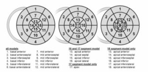
| 呼び方 | 解説 | |
|---|---|---|
| ラテラル | lateral 側壁 | |
| アンテリオール | anterior 前壁 | |
| ポステリオール | posterior 後壁 | |
| セプタール | septal 中隔 | |
| アンテロセプタル | anteroseptal 前部中隔 | |
| ミッドセプタル | midseptal 中部中隔 | |
| ポステロセプタル | posteroseptal 後部中隔 |
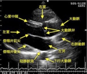
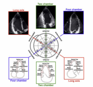
左室短軸断面像(1)大動脈弁レベル

| 男性 | 女性 | |
| 左室拡張末期径(mm):LVDd | 48±4 | 44±3 |
| 左室収縮末期径(mm):LVDs | 30±4 | 28±3 |
| 左室拡張末期容積係数(mL/m2):LV EDV | 53±11 | 49±11 |
| 左室収縮末期容積係数(mL/m2):LV ESV | 19±5 | 17±5 |
| 左室駆出率(%):LVEF | 64±5 | 66±5 |
| 左室重量係数(g/m2):LVMI | 76±16 | 70±14 |
| 左房径(mm):LAD | 32±4 | 31±3 |
| 左房容積係数(mL/m2):LAVI | 24±7 | 25±8 |
| 右室拡張末期径 (心尖部四腔断面基部)(mm):RVDd | 31±5 | 28±5 |
| 右室面積変化率(FAC, %):FAC | 44±13 | 46±11 |
| 三尖弁輪部移動距離 (TAPSE,mm):TAPSE | 24±3.5 | |
| 三尖弁輪部 s′波(cm/ 秒) | 14.1 ± 2.3 | |
| E/e′(中隔) | 7.4 ± 2.2 | 7.9 ± 2.2 |
| e′(中隔,cm/ 秒) | 10.0 ± 2.8 | 10.8 ± 3.2 |
| E/e′(側壁) | 5.5 ± 1.8 | 6.2 ± 1.8 |
| e′(側壁,cm/ 秒) | 13.5 ± 3.9 | 13.7 ± 4.1 |
LVDs: left ventricular diastolic dimension, 左室収縮期径
LV EDV: left ventricular end-diastolic volume, 左室拡張末期容積
LV ESV: left ventricular end-systolic volume, 左室収縮末期容積
BSA: body surface area, 体表面積
LV EF: left ventricular ejection fraction, 左室駆出率
SD: standard deviation, 標準偏差
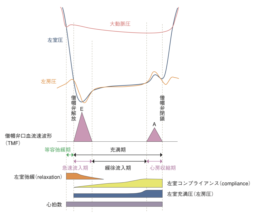
| TMF | 僧帽弁口血流速波形 | transmitral flow velocity pattern |
| E | 拡張早期波 | |
| A | 心房収縮期波 | |
| E/A | ||
| DcT | E 波減衰時間 | |
| A dur |
| RVF | 肺静脈血流速波形 | pulmonary venous flow velocity pattern |
| S2 | ||
| D | ||
| A | ||
| A dur | ||
| TDI | 組織ドプラ法 | Tissue Doppler Imaging |
| e’ | ||
| e” | ||
| E/e’ |
tethering エコー診断
左室拡大
左室壁運動障害
大動脈弁硬化
僧帽弁閉鎖不全
——————————-
心エコーの略語
PCIトップページ
MEトップページ

コメント